CT Scan Guide: Diagnostic Imaging, Preparation, and Health Information
A CT scan, or computed tomography scan, is an imaging examination that uses X-rays and computer processing to create detailed cross-sectional images of the body.
Unlike a conventional X-ray, which produces a two-dimensional image, CT can generate multiple images that can be reconstructed into different views. This allows healthcare professionals to examine bones, organs, blood vessels, soft tissues, and other structures in greater detail.
CT imaging is used in many areas of medicine, including evaluation of injuries, abdominal conditions, lung problems, vascular conditions, infections, tumors, and other medical concerns.
CT scans can be performed with or without a contrast material, depending on the clinical question and the body area being examined.
Why CT Scans Matter
CT can provide detailed anatomical information quickly, making it particularly useful in situations where rapid imaging is important.
Common applications include:
Evaluating injuries after accidents or falls
Examining the brain and head
Assessing the chest and lungs
Investigating abdominal or pelvic symptoms
Examining bones and complex fractures
Evaluating blood vessels
Identifying certain tumors or abnormal growths
Investigating unexplained bleeding
Guiding certain medical procedures
The choice of CT, MRI, ultrasound, X-ray, or another imaging method depends on the medical question, body area, urgency, and individual circumstances.
How CT Imaging Works
During a CT examination, an X-ray tube rotates around the body while detectors collect information from multiple angles.
A computer processes these measurements to create cross-sectional images, often called slices.
These images can be viewed individually or reconstructed into different planes and three-dimensional representations.
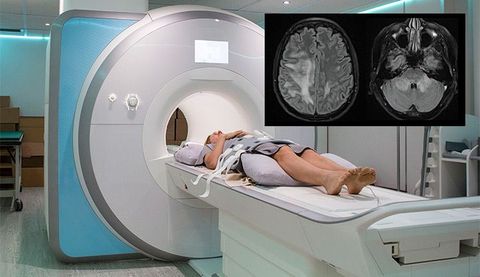
CT uses ionizing radiation, unlike MRI, which uses magnetic fields and radiofrequency energy.
Modern CT systems use technologies designed to obtain useful diagnostic images while managing radiation exposure. The amount of radiation varies according to the examination and scanning protocol.
Common Types of CT Scans
Head and brain CT
A head CT can quickly evaluate structures of the brain and skull. It may be used in certain emergency situations involving head injuries, suspected bleeding, or other neurological concerns.
Chest CT
Chest CT can provide detailed images of the lungs, airways, heart region, blood vessels, and surrounding structures.
Abdominal CT
Abdominal CT can examine organs and structures including the liver, kidneys, pancreas, spleen, intestines, and abdominal blood vessels.
Pelvic CT
Pelvic CT can evaluate structures in the lower abdomen and pelvis and may be performed alongside abdominal imaging.
CT angiography
CT angiography, or CTA, uses CT imaging and contrast material to visualize blood vessels.
CT colonography
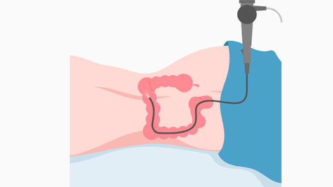
CT colonography, sometimes called virtual colonoscopy, uses CT imaging to examine the colon. It is a different examination from conventional colonoscopy and may have different preparation and follow-up requirements.
Bone CT
CT can produce detailed images of bones and may be particularly useful for complex fractures or areas that are difficult to evaluate with standard X-rays.
CT Scan With Contrast
Some CT examinations use contrast material to make particular organs, tissues, or blood vessels easier to distinguish.
Depending on the examination, contrast may be administered:
Intravenously
Orally
Through another route specified by the imaging team
Iodine-based contrast agents are commonly used for many contrast-enhanced CT examinations.
Not every CT scan requires contrast.
Before contrast administration, the healthcare team may review relevant medical information, including:
Previous reactions to contrast
Kidney function
Current medications
Relevant medical conditions
Pregnancy status when applicable
Patients should provide accurate information about previous contrast reactions and medical history before the examination.
Preparing for a CT Scan
Preparation varies according to the type of CT examination.
Some scans require little or no preparation, while others may involve dietary restrictions, fasting, oral contrast, or other instructions.
Preparation may include:
Food and drink instructions
Certain contrast-enhanced examinations may require fasting for a specified period. Patients should follow the instructions provided by the imaging facility.
Medication information
The medical team may need information about prescription medications, over-the-counter medicines, and supplements.
Contrast preparation
If oral or intravenous contrast is planned, the imaging team will provide specific instructions.
Clothing and personal items
Patients may be asked to remove jewelry, eyeglasses, or certain clothing items that could interfere with imaging.
Preparation requirements vary, so instructions from the specific imaging center should take priority.
What Happens During a CT Scan?
A typical CT examination follows several steps.
1. Registration and screening
The imaging team reviews the examination order, medical history, medications, allergies, and other relevant information.
2. Preparation
The patient may change into appropriate clothing and remove objects that could interfere with the scan.
3. Positioning
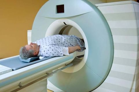
The patient lies on a motorized table that moves through the CT scanner.
4. Contrast administration
If contrast is required, it may be administered before or during the examination.
5. Image acquisition
The table moves through the scanner while X-ray measurements are collected.
6. Remaining still
Patients are generally asked to remain still. Some examinations may require briefly holding the breath.
7. Completion
The table moves out of the scanner after imaging is complete. Most routine CT examinations are relatively quick, although the total appointment time varies.
What Does a CT Scan Feel Like?
The scan itself is generally painless.
If intravenous contrast is used, some people may briefly experience sensations such as:
Warmth
Flushing
A metallic taste
A sensation similar to urination
These sensations are usually temporary, but patients should tell the medical team if they experience unexpected or concerning symptoms.
The scanner is generally less enclosed than an MRI system, which may make CT more comfortable for people who have difficulty with enclosed spaces.
CT Scan Results
CT images are typically reviewed by a radiologist, a physician specializing in medical imaging.
The radiologist prepares a report describing relevant findings. The healthcare professional who ordered the examination then interprets those findings in combination with symptoms, medical history, physical examination, and other diagnostic information.
A CT report may describe:
Normal anatomical structures
Fractures
Inflammation
Fluid collections
Abnormal masses
Bleeding
Organ changes
Blood-vessel abnormalities
Other findings requiring additional evaluation
An imaging finding does not necessarily establish a final diagnosis on its own.
Understanding CT Reports
Radiology reports can contain technical terminology.
Some common terms include:
Lesion: An area of tissue that appears different from surrounding tissue.
Nodule: A small rounded area or growth identified on imaging.
Opacity: An area that appears denser or less transparent on an image.
Calcification: An area containing calcium deposits.
Contrast enhancement: Increased visibility of tissue or a structure after contrast administration.
Impression: The radiologist's summary of the most important findings.
If a report contains unfamiliar terminology, the ordering healthcare professional can explain its significance in the context of the patient's overall medical information.
CT Scan Benefits and Limitations
CT has several important advantages:
Rapid image acquisition
Detailed cross-sectional images
Excellent evaluation of bones
Useful imaging of many internal organs
Broad availability in emergency and outpatient settings
Ability to create multiplanar and three-dimensional images
Useful for certain vascular examinations
However, CT also has limitations:
It uses ionizing radiation
Some examinations require contrast
Contrast can cause adverse reactions in some individuals
Image quality can be affected by movement
Certain findings may require additional imaging or testing
CT may provide less soft-tissue contrast than MRI for some applications
The medical team determines whether CT is appropriate based on the specific clinical question.
CT Radiation Considerations
CT uses ionizing radiation, so radiation exposure is an important consideration.
The FDA explains that medical imaging examinations involving radiation should be performed when medically appropriate, with efforts to optimize radiation exposure while obtaining the necessary diagnostic information.
Radiation dose varies substantially depending on the body region, scanner, protocol, patient size, and type of examination.
A single medically appropriate CT examination generally provides information that must be weighed against the potential risks associated with radiation exposure.
Patients should tell their healthcare team if they may be pregnant because this can affect imaging decisions and protocols.
CT Contrast Safety
Contrast material can improve visualization of certain structures, but it is not appropriate for every patient or examination.
Before contrast administration, clinicians may consider kidney function and previous contrast reactions.
Severe allergic-like reactions to modern iodinated contrast are uncommon, but reactions can occur. Imaging facilities maintain procedures for recognizing and managing adverse reactions.
Patients should report previous reactions to contrast agents before the examination.
CT vs. MRI
CT and MRI are both advanced medical-imaging technologies, but they work differently.
| Feature | CT | MRI |
|---|---|---|
| Main technology | X-rays | Magnetic field and radiofrequency energy |
| Ionizing radiation | Yes | No |
| Examination speed | Often fast | Often longer |
| Bone imaging | Excellent | Useful but different |
| Soft-tissue detail | Good | Often excellent |
| Metal-device considerations | Different safety considerations | Extensive MRI safety screening |
| Contrast | Often iodine-based when needed | Gadolinium-based agents may be used when appropriate |
Neither technology is universally better. The appropriate examination depends on the body area and medical question.
Recent Developments in CT Imaging
CT technology continues to develop through improvements in detector design, image reconstruction, scanning speed, and radiation-management techniques.
Current areas of development include:
Low-dose CT protocols
Improved image reconstruction
Faster scanning
Higher-resolution imaging
Spectral or dual-energy CT
Advanced CT angiography
AI-assisted image reconstruction and analysis
Improved motion management
More detailed quantitative imaging
These technologies can help improve image quality and workflow while supporting efforts to manage radiation exposure.
CT Screening Applications
CT is also used in selected screening programs.
One important example is low-dose CT for lung cancer screening in people who meet specific risk and age criteria.
The USPSTF recommends annual lung cancer screening with low-dose CT for adults ages 50 to 80 years who have a 20 pack-year or greater smoking history, currently smoke, or quit within the past 15 years. Screening should stop once a person has not smoked for 15 years or develops a health problem that substantially limits life expectancy or the ability to undergo curative lung cancer treatment. (uspreventiveservicestaskforce.org)
Screening recommendations can change, so eligibility should be confirmed with a healthcare professional.
Tools and Resources
Useful U.S. resources for learning about CT imaging include:
FDA — information about medical X-ray imaging and radiation safety
RadiologyInfo.org — patient-oriented information about CT examinations
American College of Radiology — imaging guidance and radiology information
National Cancer Institute — information about CT screening and cancer imaging
Radiologists and imaging specialists — professional interpretation of CT findings
FAQs
1. Does a CT scan use radiation?
Yes. CT uses X-rays, which are a form of ionizing radiation. Radiation exposure varies depending on the type of scan and imaging protocol.
2. Does every CT scan require contrast?
No. Some CT examinations are performed without contrast. Contrast is used when it may provide additional information for the specific examination.
3. How long does a CT scan take?
The actual scanning portion can be very quick, sometimes taking only a few minutes. The total appointment can take longer depending on preparation, contrast administration, and the type of examination.
4. Is a CT scan painful?
The imaging itself is generally painless. An intravenous contrast injection may cause brief sensations such as warmth or flushing.
5. Can I have a CT scan during pregnancy?
Pregnancy or possible pregnancy should always be communicated to the healthcare team before imaging. The medical team can determine whether CT is appropriate and whether an alternative examination or modified protocol is preferable.
Conclusion
CT scanning is an important diagnostic-imaging technology that uses X-rays and computer processing to create detailed cross-sectional images of the body. It is widely used for evaluating injuries, organs, blood vessels, bones, lung conditions, abdominal concerns, and many other medical questions.
Preparation depends on the examination and whether contrast material is required. Because CT uses ionizing radiation, imaging decisions should consider the clinical need and appropriate radiation-management practices.
CT results should be interpreted by qualified healthcare professionals and considered alongside symptoms, medical history, physical findings, and other diagnostic information.

Related Post...

Home Infusion Guide: Medication Delivery, Clinical Care, and Patient Resources
Updated: September 08, 2026
Read More
Cardiac Rehabilitation Guide: Heart Recovery, Exercise, and Care Planning
Updated: September 08, 2026
Read More
Genetic Testing Guide: DNA Screening, Medical Insights, and Health Information
Updated: September 08, 2026
Read More